IHC Protocol
Arrows indicate positive SenTraGor staining in Senescent Hodgkin and Reed Sternberg cells of a primary human Hodgkin’s Lymphoma. Chromogen: DAB, Counterstain: Hematoxylin
Read more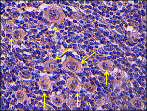
Arrows indicate positive SenTraGor staining in Senescent Hodgkin and Reed Sternberg cells of a primary human Hodgkin’s Lymphoma. Chromogen: DAB, Counterstain: Hematoxylin
Read more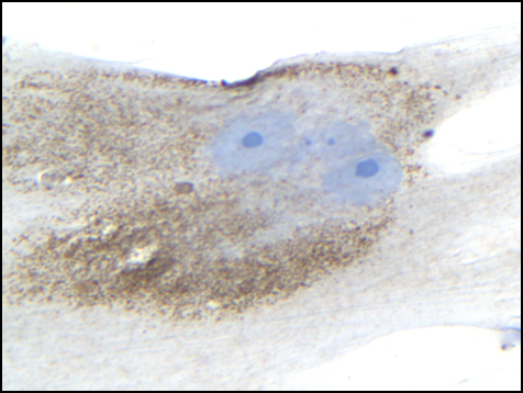
Positive SenTraGor staining in senescent and in late passage human diploid lung fibroblasts (DLFs), undergoing replicative senescence. Chromogen: DAB, Counterstain: Hematoxylin
Read more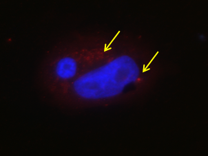
Arrows indicate positive SenTraGor staining (Texas Red) in senescent U2OS Cdt1 Tet-ON cells. Counterstain: DAPI
Read more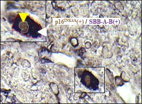
Representative images from double-staining experiments in mouse models (K-ras V12-induced lung adenoma), showing nuclear p16INK4Aorp21WAF1/Cip1expression (DAB IHC-brown color: yellow arrowheads) in senescent cells that are concurrently positive with the GL13 compound, visualized with the BCIP/NBT chromogenic hybrid Histo-IHC reaction (dark blue perinuclear and cytoplasmic color: white arrowheads; red dashed line: cell perimeter; white dashed line: nuclear perimeter.
Read more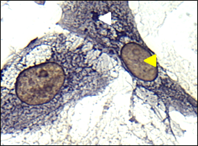
Representative images from double-staining experiments in induced Saos2-p21WAF1/Cip1 cells. DAB IHC-brown color for p16INK4A and p21WAF1/Cip1, respectively (yellow arrowheads) in senescent cells concurrently positive with Sentragor, visualized with the BCIP/NBT chromogenic hybrid Histo-IHC reaction (dark blue perinuclear and cytoplasmic color).
Read more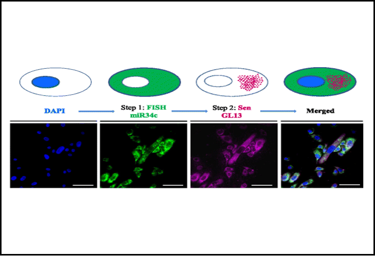
In situ detection of miR34c in senescent HBEC CDC6 Tet-ON cells. FISH of miR34c visualized as green emission in the cytoplasm, using TSA plus Fluorescein (emitting at 518 nm) and concurrent Sentragor staining, visualized at far red spectra as granules in the cytoplasm employing Alexa Fluor goat-anti-mouse.
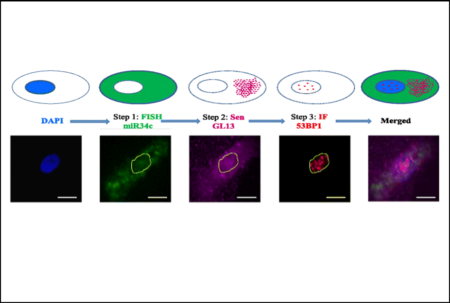
Co-detection of miR34c and 53BP1 in senescent HBEC CDC6 Tet-ON cells. miR34c FISH, visualized as green emission in the cytoplasm, using TSA plus Fluorescein; Sentragor staining, visualized as granules in the cytoplasm employing Alexa Fluor goat-anti-mouse and 53BP1 IF, visualized as red foci in the nucleus, employing Alexa Fluor goat-anti-mouse.
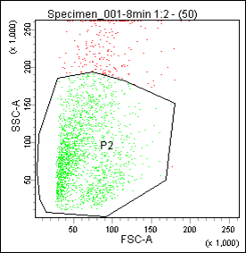
Li-Fraumeni senescent cells (Galanos et al. Nat Cell Biol. 2016 Jul;18(7):777-89) were stained with GL13 (SenTraGor) and analysed with flow cytometry. P2, viable cells; P3, GL13-positive cells. Vassilis Gorgoulis’ lab in collaboration with Ourania Tsitsilonis’ lab.
Read more